Detection of Scrotal Pathology with Ultrasound
Introduction
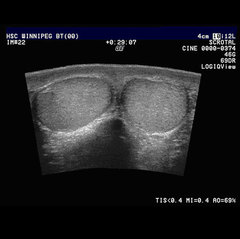
This program will focus on the sonographic evaluation of the scrotum and scrotal pathology. Sonography plays an important role in evaluating testicular size, differentiating between intratesticular or extratesticular abnormalities causing scrotal enlargement or a palpable mass, finding an occult (concealed) neoplasm, evaluating the condition of the testicle in cases of trauma or infection, and determining the presence or absence of a varicocele in an infertility workup. The majority of extratesticular masses are benign, but the majority of intratesticular masses are malignant. It is also important for the sonographer to describe the appearance of the mass, to determine if it is cystic or solid, well-defined or irregular.
Clinically a scrotal mass may be present or found on physical examination. Sometimes the mass is accompanied by pain as hemorrhage into the tumor can produce pain and tenderness. The clinical findings, pertinent lab data, and sonographic evaluation will be presented for the major scrotal pathologies seen by ultrasound.
Sandra L. Hagen-Ansert, M.S., RDMS, RDCS (F)SDMS
Ultrasound Education Specialist and Clinical Consultant
Charleston, South Carolina